Structure and function of fatty acid synthases

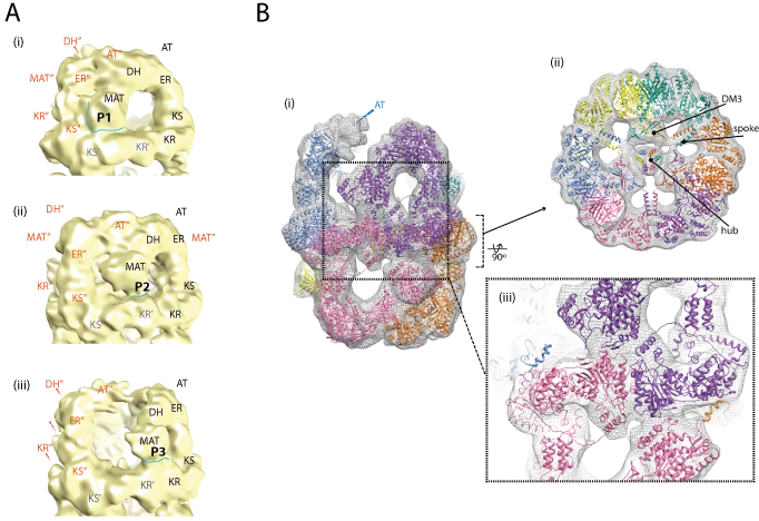
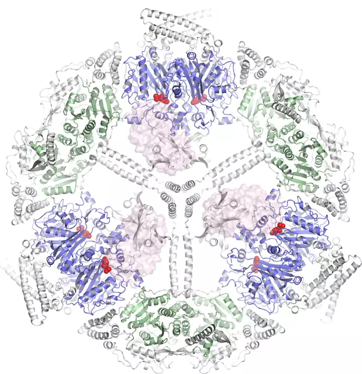
Important advances in the characterization of fatty acid synthase type I (FAS I) multienzyme complexes have been made during the last years by reports on X-ray crystallographic and cryo-electron microscopic (cryo-EM) structures of the bacterial, fungal and mammalian megacomplexes. In a program to structurally characterize FAS I systems, we were able to significantly contribute to this new understanding. Specifically, we structurally characterized the fungal and the bacterial FAS I, and shed light onto the acyl carrier protein (ACP) mediated substrate shuttling as well as the conformation dynamics as important inherent property of FAS I systems.

Fatty acids are carboxylic acids consisting of a polar carboxyl head group and a hydrophobic carbon tail. According to their nature, fatty acids are found between lipid and water with the carboxyl group soluble and exposed in water and the aliphatic chain elongated into the lipid phase. Most fatty acids are, however, not existing as a free fatty acid, but via ester or amide linkages as a substructure of lipids or proteins. While there is a huge variety of naturally occurring fatty acids known, a single organism uses only a few of them. In S. cerevisiae C16 (palmitic) and C18 acids (stearic) predominate. Fatty acid synthesis is performed in type I and type II systems. While in type II systems every catalytic function is provided by a separate enzyme, in type I systems, several catalytic centers are placed in a single polypeptide chain. FAS I are large multimeric proteins and generally show complex architectures.

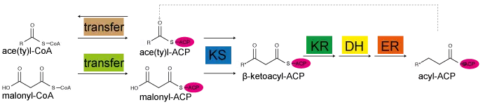
Figure. Fatty acid cycle.

Fatty acid synthesis performed in bacterial and fungal type I systems. A ketoacyl synthase (KS) domain condenses acetyl with malonyl to form β-ketobutyryl. This compound is reduced by a NADPH-dependent ketoacyl reductase (KR) to β-hydroxyacyl, dehydrated by a dehydratase (DH) to produce enoyl, and finally reduced by an enoyl reductase (ER) to the saturated acyl-product. In all these steps, the intermediates are held by ACP. Transferases load and unload ACP.

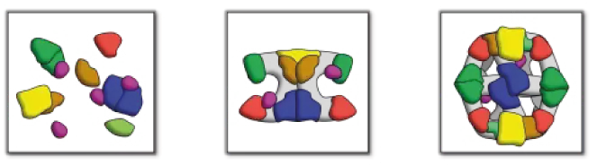
Figure. Fatty acid systems. Schematic representation of type I and type II systems. The left panel shows the separate proteins of FAS II, while complex architectural arrangements of the mammalian FAS I and fungal/bacterial FAS I are abstracted in the middle and the right panel, respectively.

Modular polyketide synthases (PKSs): a new model fits all?

Perspectives on the evolution, assembly and conformational dynamics of fatty acid synthase type I (FAS I) systems.


The structure of the 1.9 MDa Mycobacterium tuberculosis FAS I

The bacterial FAS I is 2.0 MDa large multienzymatic molecular machine. Although considerably reduced in molecular weight compared to as fungal FAS I, the evolutionary relationship to fungal FAS I was evident from sequence comparison. In a recent work on the electron microscopic characterization, we revealed that, although overall homologous to rigid fungal FAS I, the bacterial protein is of high conformational variability allowing dynamic domain reorganizations during fatty acid synthesis

Figure. Domain arrangements in M. tuberculosis FAS I and molecular model of state P3. (A) MAT positions P1 (i), P2 (ii) and P3 (iii). In EM maps 1-3, three states could be identified with characteristic positioning of domains. As in this states MAT is displaced largest, the states are denoted P1, P2 and P3 according to MAT positions. The domain labels KS, KR, DH, ER, AT and MAT in each chain are distinctly colored and denoted with a superscript. (B) Homology model of M. tuberculosis FAS map 1 with state P3 in the upper dome. Chains are highlighted by different coloring.

Further information: In addition to the broad impact of our work on the understanding of bacterial FAS I in general, the presented data are also of specific importance. M. tuberculosis is the causative agent of tuberculosis (TB). Its FAS I is responsible for de novo fatty acid synthesis, forming precursors for mycolic acids that build up the impermeable, wax-like cell wall and make M. tuberculosis impervious to medical drugs. The relevance of M. tuberculosis FAS as drug target is documented by pyrazinamide, which has been identified as specifically targeting M. tuberculosis FAS I and is used in current antibiotic therapy.

We consider our data as highly relevant for anti-tuberculosis initiatives. We demonstrated that M. tuberculosis FAS I can be recombinantly expressed in Escherichia coli, which will facilitate future functional and structural investigations on this highly demanded drug target. Moreover, our data potentially contribute in establishing M. tuberculosis FAS I as a target for rational drug design.

Structure and conformational variability of the mycobacterium tuberculosis fatty acid synthase multienzyme complex.


The structure of the 2.6 MDa Saccharomyces cerevisiae FAS I

The 2.6 MDa fungal FAS I consists of two dome-shaped reaction chambers formed by six β-chains, each 230 kDa in molecular weight, and an equatorial wheel-like structure made-up by six α-chains, each 210 kDa. The catalytic domains are interspersed by a number of scaffolding domains, which allow the formation of the complex barrel-shaped architecture, in which the catalytic domains are directing towards the hollow reaction chambers.

In FAS I, ACP has a key role. It takes up substrates and carriers them from enzymatic domain to enzymatic domain. S. cerevisiae FAS I proved to be an excellent system studying substrate shuttling in more detail. In collaboration with the Max-Planck-Institute of Biophysics, we were able to give direct insight finding ACP docked at active sites. This allowed us getting direct feedback on ACP docking, likely coupled to reaction rates, and allowed us to characterized key properties in a molecular dynamics modeling approach

Direct structural insight into the substrate-shuttling mechanism of yeast fatty acid synthase by electron cryomicroscopy.

Multimeric options for the auto-activation of the Saccharomyces cerevisiae FAS type I megasynthase.